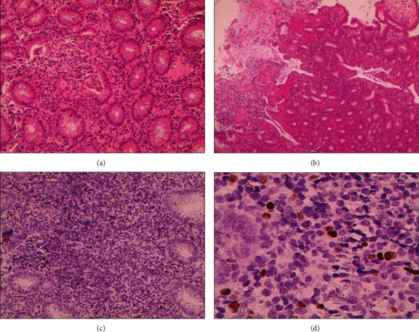
Figure 2.

Histopathology of ulcerative colitis in patients with intestinal EBV infection: (a) magnification 200x; (b) 40x; HE staining. The mucosa of large intestine showed active chronic colitis, with fossa inflammation, fossa thickening, and local ulcer formation. (c) 200x; (d) 400x. EBER demonstrated EBV-positive lymphocytes of 40/HPF.
