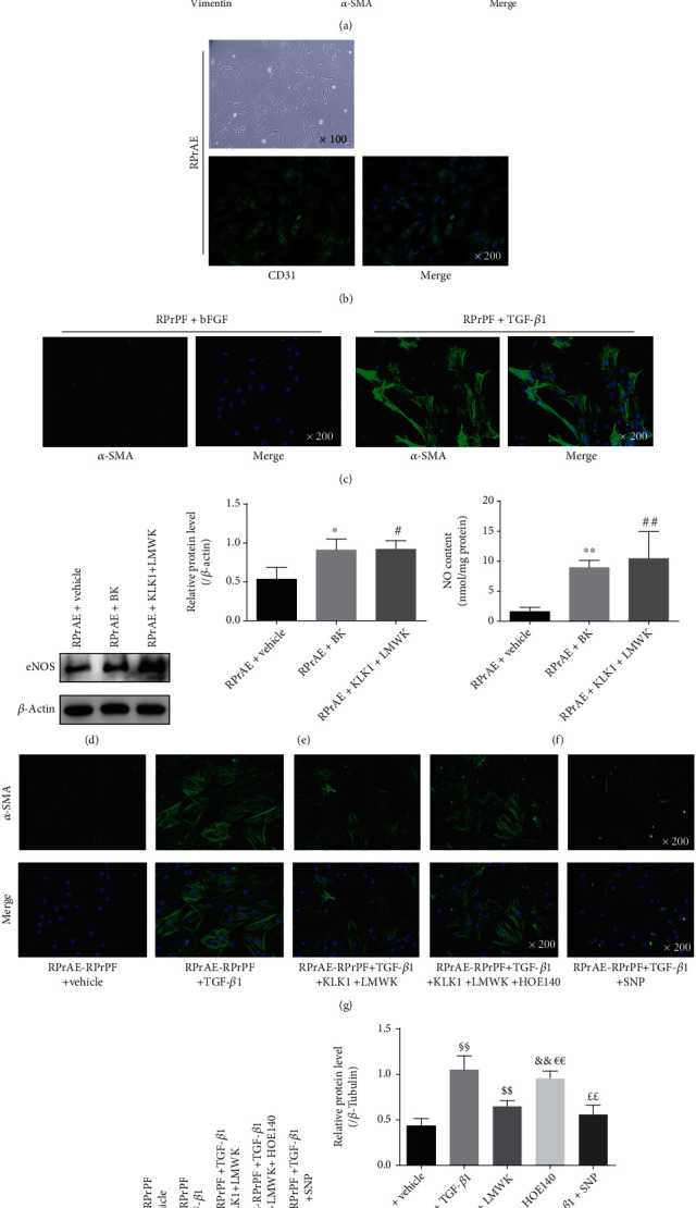
Figure 2.

KLK1 could inhibit fibroblast-myofibroblast transdifferentiation induced by TGF-β1 in RPrAE-RPrPF coculture system. (a) The morphology of RPrPF (magnification ×100) and the verification of RPrPF through the expression of E-Cadherin (red), vimentin (red), and α-SMA (green) (magnification ×200). (b) The morphology of RPrAE (magnification ×100) and the verification of RPrAE through the expression of CD31 (green) (magnification ×200). (c) The fibroblast-myofibroblast transdifferentiation induced by TGF-β1 in RPrPF (α-SMA, green; magnification ×200). (d, e) Protein expressions of eNOS normalized to β-actin in RPrAE under KLK1, LWMK, or BK. (f) NO content in RPrAE under KLK1, LWMK, or BK. (g) Representative IF photos of α-SMA (green) expression level changes at the administration of TGF-β1, KLK1, LWMK, HOE140, and SNP in RPrAE-RPrPF coculture system. (h, i) Protein expressions of α-SMA normalized to β-Tubulin in RPrAE-RPrPF coculture system by Western blot. Each bar represents mean ± SD of 3 independent repeated experiment. ∗P < 0.05, ∗∗P < 0.01 (RPrAE + BK vs. RPrAE + vehicle). #P < 0.05, ##P < 0.01 (RPrAE + KLK1 + LMWK vs. RPrAE + vehicle). §§P < 0.01 (+ TGF-β1 vs. + vehicle). $$P < 0.01 (+ TGF-β1 + KLK1 + LMWK vs. + TGF-β1). &&P < 0.01 (+ TGF-β1 + KLK1 + LMWK + HOE140 vs. + vehicle). €€P < 0.01 (+ TGF-β1 + KLK1 + LMWK + HOE140 vs. + TGF-β1 + KLK1 + LMWK). ££P < 0.01 (+ TGF-β1 + SNP vs. + TGF-β1).
