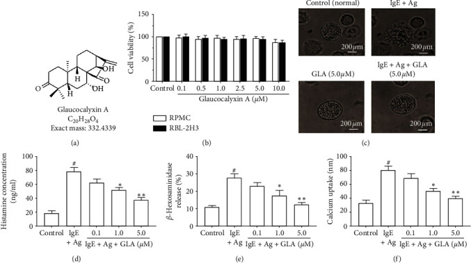
Figure 2.

Effect of GLA on cell viability and degranulation in RPMCs and RBL-2H3 cells. (a) Chemical structure of GLA. (b) Measurement of cell viability using MTT assay. RPMCs were divided into control, IgE + Ag (sensitized with 50 ng/mL anti-DNP IgE for 6 h and challenged with 100 ng/ml DNP-HSA) and IgE + Ag + GLA (sensitized with 50 ng/mL anti-DNP IgE for 6 h treated with 5 μg/mL GLA and then challenged with 100 ng/ml DNP-HSA). (c) Morphology changes of degranulation of RPMCs (magnification, ×1,000). (d) Histamine concentration. (e) β-Hexosaminidase release. (f) Calcium uptake. All data represent mean ± SEM (n = 3). Compared with the control group, #P < 0.05. Compared with the IgE + Ag group, ∗P < 0.05 and ∗∗P < 0.01.
