Figure 2.

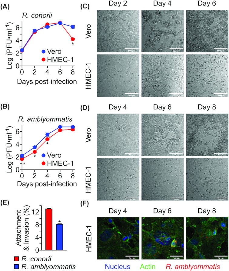
Rickettsia amblyommatis is defective for attachment to host endothelial cells and replicates at a slower rate. (A)Rickettsia conorii strain Malish 7 and (B)R. amblyommatis strain GAT-30V replications in Vero and HMEC-1 cells were quantified with the plaque assay (mean ± SEM, N = 3). Representative differential interference contrast (DIC) microscopic images of Vero and HMEC-1 cells infected with (C)R. conorii or (D)R. amblyommatis on 2, 4, 6 and 8 days post-inoculation (scale bar, 500 µm). (E)Rickettsia conorii strain Malish 7 and R. amblyommatis strain GAT-30V attachment and invasion into HMEC-1 cells were quantified with the plaque assay on Vero cells (mean ± SEM, N = 3). (F) Representative confocal microscopic images of intracellular R. amblyommatis in HMEC-1 cells (scale bar, 30 µm). *, P < 0.05. Data are representative of two independent experiments.
