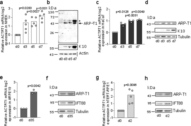
Fig. 1. ARP-T1 is expressed during epidermal and epithelial differentiation.
a, c, e, g mRNA expression of ACTRT1 during differentiation of keratinocytes, NHEK (a N = 5) and HaCaT (c N = 3), and epithelial cells, ARPE19 (e N = 3) and hTERT-RPE1 (g N = 5). Data are presented as means of the fold change compared to the value of undifferentiated samples. Each open circle represents one independent experiment. b, d, f, h Representative images of ARP-T1 expression during differentiation of keratinocytes, NHEK (b) and HaCaT (d), and epithelial cells, ARPE19 (f) and hTERT-RPE1 (h). ARP-T1 was detected using guinea pig anti-ARP-T1 antisera (b) or mouse antibody (d, f, h). * indicates polymers of ARP-T1 confirmed by mass spectrometry analysis. Keratin 10 and IFT88 were used as markers of cell differentiation in keratinocytes and epithelial cells, respectively, actin and tubulin as loading controls.

