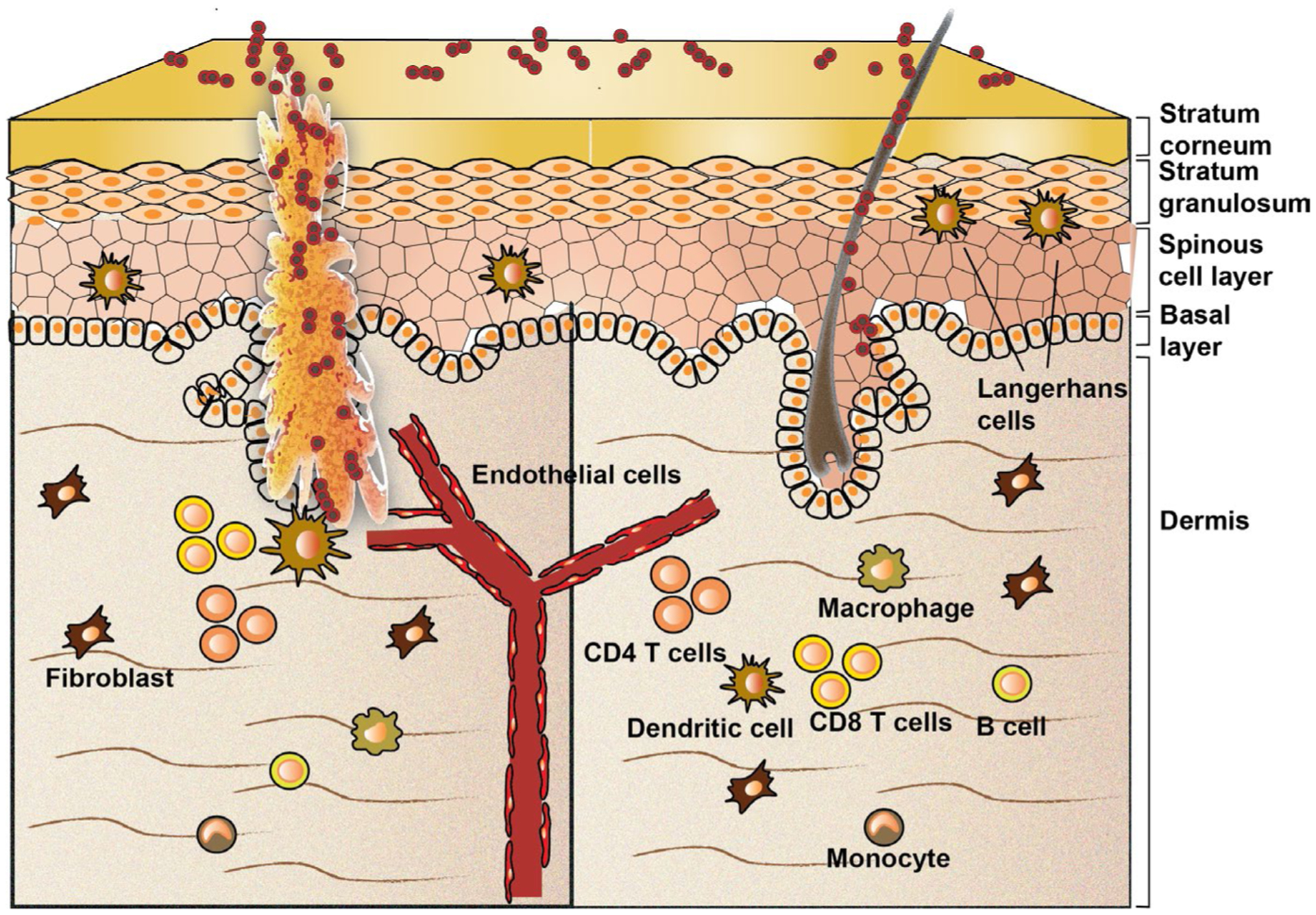
FIGURE 3.

Potential antigen-presenting cells (APCs) that are present in the skin. When the skin barrier is disrupted, Staphylococcus aureus can colonize the open wound and become pathogenic. Professional APCs, such as macrophages, Langerhans cells, dendritic cells and B cells, can internalize and process S. aureus peptide fragments in lysosomes. Generated peptide fragments can be loaded onto major histocompatibility complex (MHC) class II molecules. Non-professional APCs, such as keratinocytes, fibroblasts and endothelial cells, also have the capacity to internalize and process peptide fragments of S. aureus. These cells activate their autophagy machinery as their lysosomal degradation capacity is limited. APC rely on Perforin-2–mediated kill of intracellular pathogens
