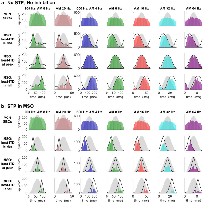
Fig. 4.
MODEL 1: AMBB-cycle spike-rate histograms of SBCs and MSO neuron for AMBBs centred at CFs 200 Hz (leftmost 2 columns) and 600 Hz (rightmost 5 columns): (a) with no inhibition and no STP; (b) with STP in the MSO. Definitions: grey silhouettes: AM envelope; black lines: normalised static-IPD functions for a single amplitude-modulated carrier frequency at CF. The model MSO neuron has a best ITD of zero: ‘best-ITD in rise’ denotes start-IPD = −90° (270°); ‘best-ITD at peak’ denotes start-IPD = −180° (180°); and ‘best-ITD in fall’ denotes start-IPD = −270° (90°)

