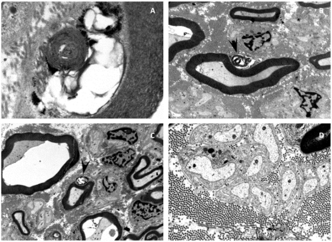
Figure 1.
Electron microscopy of the sural nerve of patient I. Electron microscopy showed neurodegenerative processes and inclusions in the Schwann cells of myelinated axons of the sural nerve (A–D). Some of these inclusions can be identified as myelin-like figures with concentric lamellar material and periodicity of about 10 nm (A). The inclusions are often mixed with glycogen-like granules (C). Membranous vacuolized and optically empty bodies, as well as isolated irregularly contoured electron-dense and sharply delimitable “tufa”-like inclusions (B,C). These inclusions lead to the suspicion of a lysosomal storage disease.

