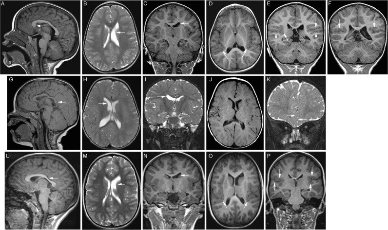
Fig. 2. Brain MRI demonstrates features of classic tubulinopathies with or without MCD.
A–F MR images from Individual 1 at 20 months of age. A Midline sagittal T1 FLAIR MR image reveals deficiency of the rostrum of the corpus callosum. B Axial T2 and (C) coronal T1 MPRAGE MR images demonstrate an asymmetrically smaller, foreshortened body of the left caudate nucleus (white arrows) with mild prominence and dysmorphism of the ipsilateral lateral ventricle. D Axial T1 FLAIR MR image (5 mm/2.25 mm gap thickness) demonstrates thickening and irregularity of the posterior perisylvian cortex (white arrows), most pronounced on the right. E, F Coronal T1 MPRAGE MR images (0.9 mm/0 mm gap thickness) confirm bilateral perisylvian polymicrogyria (white arrows). G–I MR images from Individual 2 at 13 months of age. G 1.5T Siemens Symphony midline sagittal T1 MR image (4 mm/0.8 mm gap thickness) reveals mild thinning of the body and splenium (arrow) of the corpus callosum. H Axial (4 mm/0.8 mm gap) and (I) coronal (2 mm/ 0 mm gap) turbo spin echo T2 weighted MR images show asymmetric lateral ventricles and caudate nuclei, with the body of the right caudate nucleus appearing slightly smaller than the left (arrow). J Axial spin echo 1.5 T Siemens Symphony T1 MR image (4 mm/ 0.8 mm gap) demonstrating the perisylvian cortex. The cortical architecture is not well assessed for MCD due to a combination of technical parameters and age-appropriate incompletely myelinated subcortical white matter. K Anterior coronal (2 mm/ 0 mm gap) turbo spin echo T2 weighted MR image shows interdigitating frontal lobes (arrows) attributed to deficiency of the falx cerebri. L–P MR images from Individual 3 at 7 years of age. L Midline reconstructed sagittal T1 MPRAGE MR image (0.8 mm/ 0 mm interslice gap) reveals accentuation of the usual thinning of the posterior body of the corpus callosum (arrow). M Axial T2 (4 mm/1.2 mm gap) and N coronal T1 MPRAGE (1.2 mm/0 mm gap) MR images show asymmetric lateral ventricles and caudate nuclei, with the body of the left caudate nucleus appearing smaller than the right (arrow). O Reformatted axial (0.8 mm thickness) and (P) direct coronal (1.2 mm/ 0 mm gap) T1 3D MPRAGE MR images demonstrate irregular, thickened perisylvian cortex, more pronounced on the right, consistent with polymicrogyria (arrows).

