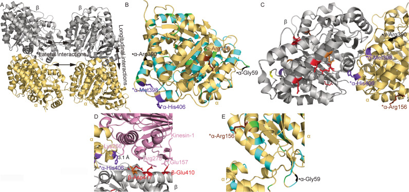
Fig. 3. Structural modeling of tubulinopathy variants.
Structures were obtained from PDB ID: 3JAK (A–C, E) and PDB ID: 4HNA (D). A α and β tubulin monomers within a portion of a microtubule. Representative lateral and longitudinal interfaces are indicated. B α tubulin monomer with TUBA1A tubulinopathy-associated residues colored. C α and β tubulin monomers at the heterodimerization interface. Colored beta tubulin residues are associated with substitutions in TUBB3 and/or TUBB2B. For simplicity, residues associated with isolated MCD or MCD with involvement of cranial nerves other than CN3 are omitted in this view. D α and β tubulin complexed with kinesin-1. Colored beta tubulin residues are associated with substitutions in TUBB3 and/or TUBB2B. For simplicity, residues associated with isolated MCD or MCD with involvement of cranial nerves other than CN3 are omitted in this view. Polar interactions between kinesin and alpha or beta tubulin residues are shown with dashed lines. Distance between kinesin-Lys256 and α-His406 is shown. E Lateral interface between α tubulin residues in adjacent microtubule protofilaments. Key: α tubulin (gold); β tubulin (silver); kinesin-1 (pink); residues associated with CFEOM but not MCD (red); residues associated with CFEOM+MCD (purple); residues associated with MCD in the absence of CFEOM (cyan); residues associated with CFEOM ± MCD (orange); residues associated with CFEOM in the absence of MCD, or associated with MCD in the absence of CFEOM depending on substitution (brown); residues associated with MCD ± CFEOM (yellow); previously reported residues associated with putative CFEOM in the absence of MCD (black; [50]); reported residues associated with MCD and anomalies of cranial nerves other than CN III (green; [25]). *residues reported for the first time in this work (Arg156, Met398, and His406). ●-Previously reported residues associated with putative CN3 phenotypes [50].

