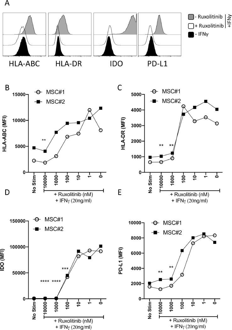
Figure 2. IFNγ induced HLA-ABC, HLA-DR, PDL-1 and IDO expression in MSCs are suppressed by Ruxolitinib.
MSCs were treated with contemporaneous Ruxolitinib and IFNγ (20ng/mL) for 48 hours. Cells were stained for flow cytometry analysis using antibodies against MHC I (HLA ABC), MHC II (HLA-DR), IDO and PDL1. (A) Representative histograms are shown. Grey and white histograms represent – and + Ruxolitinib in the presence of IFNγ. Black histogram represents unstimulated control. Dose dependent effect of Ruxolitinib on the mean fluorescent intensity (MFI) of (B) MHC I (HLA ABC), (C) MHC II (HLA-DR), (D) IDO and (E) PDL1 is shown on two independent MSC donors. Similar results were obtained in a repeat experiment. Two-Way ANOVA multiple comparison test was performed between 0 nM and other Ruxolitinib concentrations. **, ***, **** represents P ≤ 0.01, P ≤ 0.001 and P ≤ 0.0001 respectively. Cumulative statistical significance with both MSC donors is shown.

