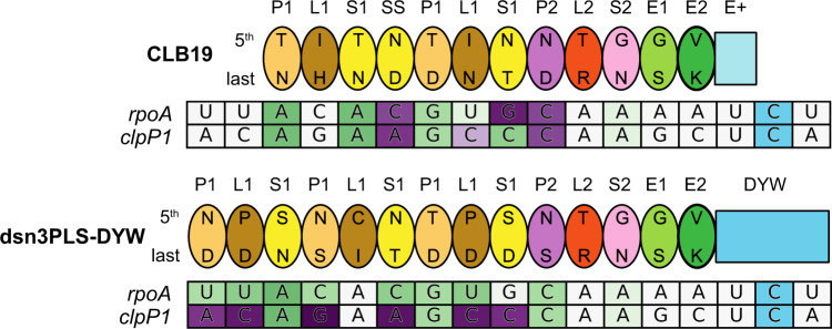
Fig. 1. Schematic representation of CLB19 and dsnPLS-DYW bound to their target sites.
The proteins are represented by ovals for each PPR motif including the 5th and last specificity-determining amino acids. The target sites are coloured according to the predicted favourability of the alignment at each position, from favoured (green) to neutral (white) to disfavoured (purple). These values are taken from ref. 26. The C at the editing site is shaded in blue.

