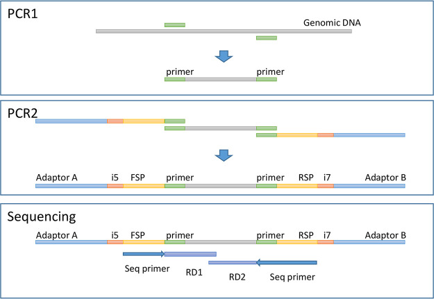
FIG 1.
(Top) PCR1. Shown is a diagram of the PCR of the gene target area with genomic DNA as the template. Genomic template DNA was subjected to PCR-based amplification with one primer set targeting prokaryotes and three primer sets targeting eukaryotes. Each PCR was run in parallel. (Center) PCR2. Shown is a diagram of the attachment of the required elements to amplicons for MiSeq sequencing. The products from PCR1 were used as templates for the adaptor PCR, where adaptors, bar codes, and sequencing primer-binding sites were added. This was performed in parallel for each of the four primer sets. (Bottom) Sequencing. Shown is a diagram of the regions sequenced by MiSeq as RD1 and RD2. See the text for details.

