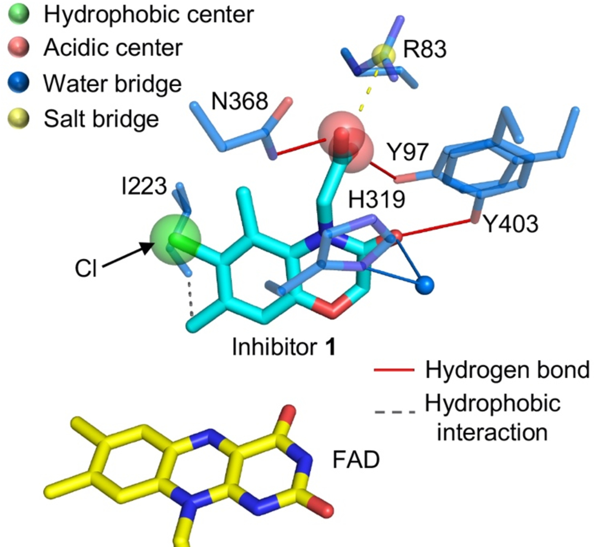
Figure 3. Structure basis of brain penetrating KMO inhibitors.

The interaction between compound 1 and Pseudomonas fluorescens KMO [30] (Structure file acquired from Protein Data Bank, 6FP1, https://www.rcsb.org). Key active site residues are shown in atom coloured sticks, carbons of KMO in blue, inhibitor 1 in cyan and FAD in yellow.
