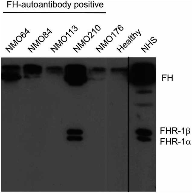
Figure 3.

Detection of FH–autoantibody complexes. Western blot analysis of the IgG fractions for FH/FHR-1 – IgG complexes. 10 µl of serum samples were incubated with Protein G beads. The bound proteins, eluted with SDS-sample buffer, were run on 10% SDS-PAGE, transferred to nitrocellulose membrane, and the blot was developed with monoclonal anti-FH recognizing also FHR-1 (mAb C18). FH is detected in the FH-autoantibody positive NMOSD patients (NMO64, NMO84, NMO210), but not in the FH-autoantibody negative (NMO176) or healthy control sample. In addition, FHR-1 is also seen in the NMOSD sample with the highest autoantibody titer (NMO210). Normal human serum (NHS) was run as a control. The blot is representative of three experiments.
