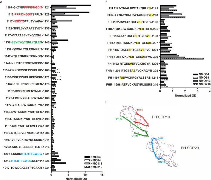
Figure 7.
Linear epitope mapping of the FH autoantibodies. Overlapping 15-mer solid phase peptides (A) covering the 19-20 domains of FH and (B) containing the FH S1191L and V1197A FHR-1 specific amino acid exchanges (indicated by yellow highlighting) were incubated with patients’ sera. Autoantibody binding was detected using HRP-conjugated anti-human IgG, and is expressed as ratio of ODsample/ODmin, where ODsample is the mean of duplicate OD values of the patients’ samples, while ODmin represents the mean antibody binding to the negative control HSP480-489 peptide. On the y axis the initial and final amino acid of each tested peptide is displayed with the single-letter amino acid sequence indicated in between. (C) The schematic picture of the FH C-terminal domains shows the identified epitopes highlighted in red (1112-1121), green (1132-1146) and blue (1212-1221), corresponding to the color codes of the one-letter amino codes in A.

