Fig. 2.
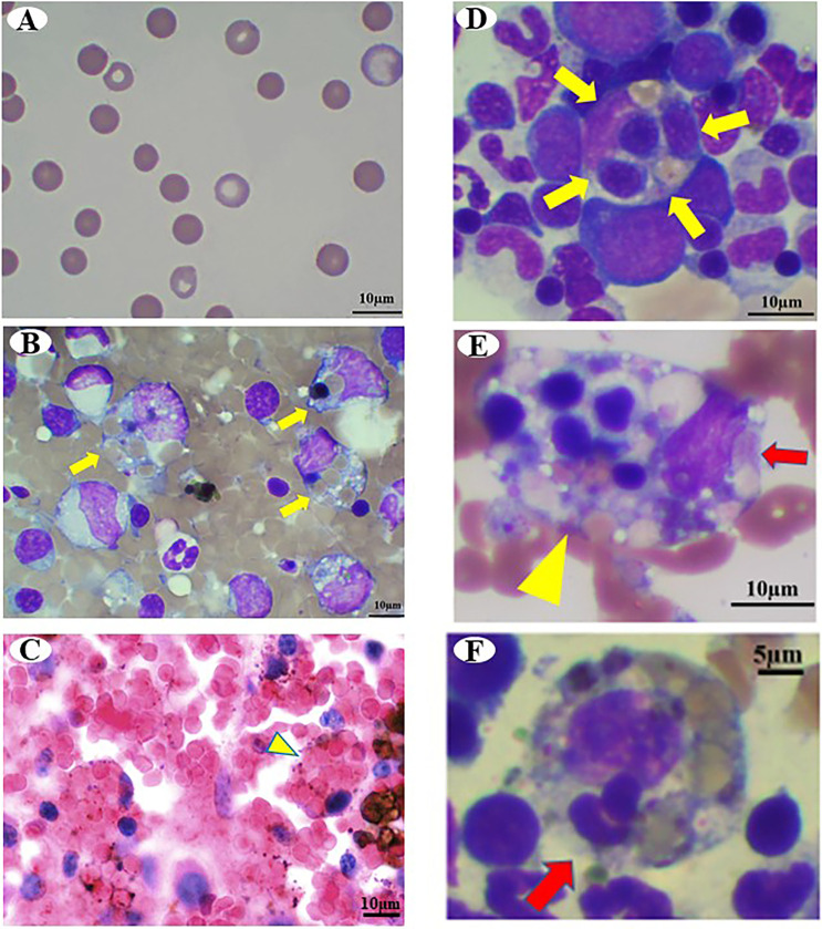
Photomicrographs of Case 1. (A) Peripheral blood smear shows increased spherocytes on day 1. Wright Giemsa stain, Bar=10 µm. (B) Cytology of the spleen. Yellow arrows indicate macrophage phagocytosis. Wright Giemsa stain, Bar=10 µm. (C) Histology of the spleen. Yellow arrowheads indicate erythrophagocytic macrophages. Hematoxylin and eosin (H&E) stain, Bar=10 µm. (D–F) Cytology of the bone marrow. (D) Yellow arrows indicate hemophagocytosis of erythroid precursors and mature erythrocytes. Wright Giemsa stain, Bar=10 µm. (E) Yellow arrowhead indicates hemophagocytic macrophage. Red arrows indicate platelets phagocyted by macrophages. Wright Giemsa stain, Bar=10 µm. (F) Red arrow indicates leukocyte phagocyted by macrophages. Wright Giemsa stain, Bar=5 µm.

