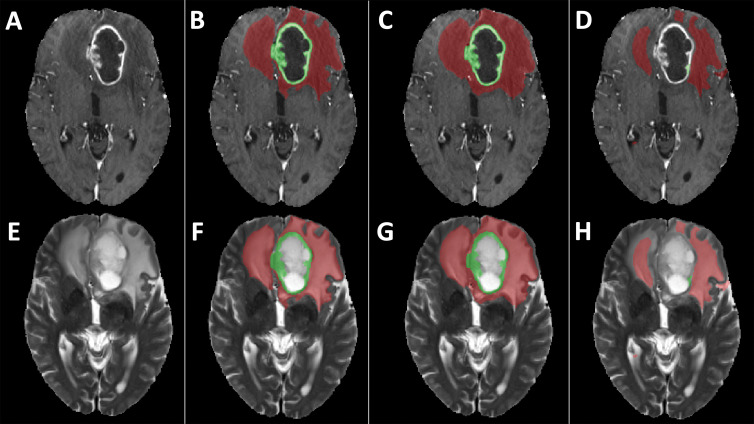
Figure 4:
Examples of segmentation masks. A–D, Postcontrast T1-weighted images and, E–H, T2-weighted images of glioblastoma from Multimodal Brain Tumor Segmentation Challenge test set. Segmentation masks of contrast-enhanced area (green) and T2 hyperintense area (red) obtained, B, F, with segmentation model using only original scans as inputs (scenario 1), C, G, using images generated with generative adversarial networks (GANs) (scenario 4), and, D, H, using copies of other scans (scenario 7). Use of only empty scans (scenario 10) failed to produce any masks (not shown). When compared with ground truth segmentations (B, F), dice similarity coefficient for whole segmentation masks obtained with GANs (C, G) was 0.88, whereas dice similarity coefficient obtained with copies of other available scans (D, H) was 0.56.

