Figure S2.
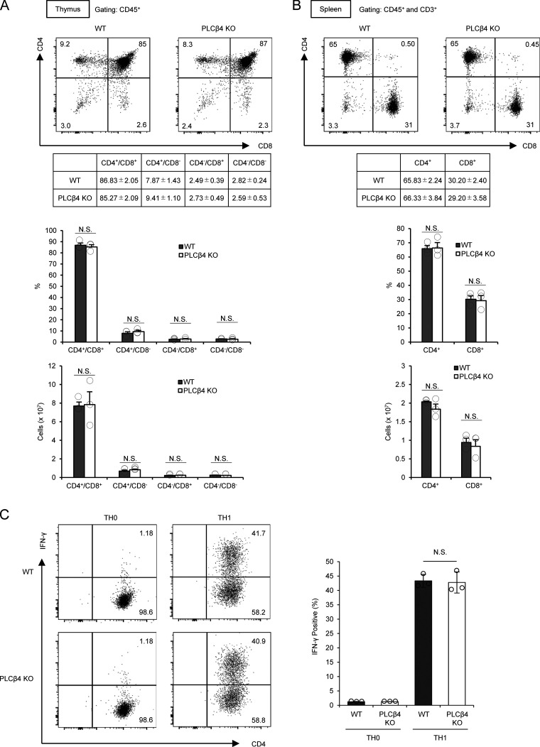
PLCβ4-deficient mice show intact T cell development and Th1 differentiation. (A) Percentages and cell numbers of CD4−/CD8−, CD4+/CD8−, CD4−/CD8+, and CD4+/CD8+ in the thymocytes from WT or PLCβ4-deficient mice were analyzed by flow cytometry. (B) Percentages and cell numbers of CD4+ and CD8+ T cells in the splenocytes from WT or PLCβ4-deficient mice were analyzed by flow cytometry. (C) Naive CD4+ T cells from WT or PLCβ4-deficient mice were cultured under Th1-polarizing conditions for 3 d. Frequencies of IFN-γ producers were determined by intracellular staining using flow cytometry. Indicated values are means ± SD of three biological replicates (A–C). N.S., nonsignificant.

