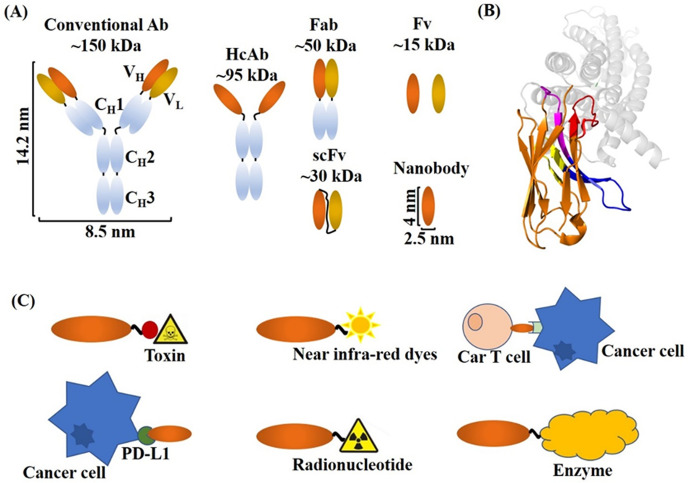
Figure 1.
Depiction of Nb structure and their applications in cancer therapy and diagnosis. (A) Schematic representation of different Ab formats. Conventional Abs consist of two light chains and two heavy chains. HcAbs consist of two identical heavy chains only. Nb is the smallest naturally occurring antigen binding fragment. (B) A crystal structure of an Nb binding its antigen G protein-coupled receptor (GPCR). GPCR is shown in gray, the FR regions (except FR2) are in orange, the FR2 region consisting of featured hydrophilic amino acids, CDR1, CDR2, and the prolonged CDR3 regions are shown in blue, magenta, yellow, and red, respectively (PDB ID: 4XT1). (C) Schematic representations of the applications of Nb conjugates in cancer therapy.

