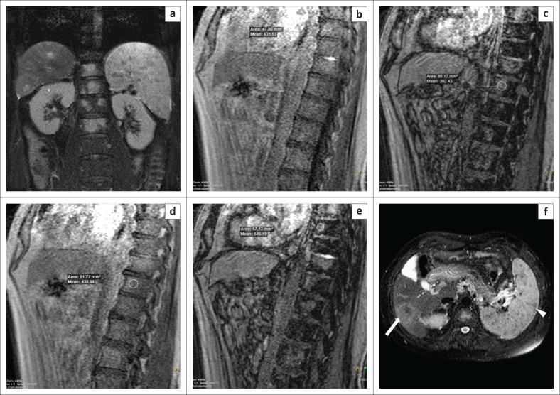
FIGURE 3.
Vertebral metastases. A 67-year-old man complained of back pain for 1 month. Magnetic resonance imaging showed multiple areas of abnormal signal within the thoracolumbar vertebrae, which were hyperintense on coronal T2W spectral attenuated inversion recovery (a) images. By comparing the in-phase (b, d) and opposed-phase (c, e) images, it was determined that the decrease in signal intensity on the opposed-phase images was 13% and 10% (<20%) at two different vertebral levels, which indicated the probability of metastases. Upon screening the abdomen, it was observed that the patient also had multiple lesions in the spleen (white arrowhead) and a lesion in the liver within segment VI (white arrow), as seen on the axial T2W spectral attenuated inversion recovery image of the abdomen (f). We therefore concluded that the vertebral lesions were metastases. However, the patient did not undergo a biopsy.

