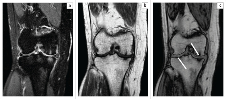
FIGURE 5.
Degenerative changes. A 49-year-old female presented with a history of chronic right knee pain. The Proton-Density-Weighted spectral attenuated inversion recovery image (a) showed areas of subchondral hyperintensity, which was suggestive of marrow oedema. On the T1W (b) image, subchondral marrow hypointensity was observed. On the opposed-phase image (c), thinning of the cartilage (white arrows) at the site of the subchondral signal change was observed, which was consistent with changes of osteoarthritis.

