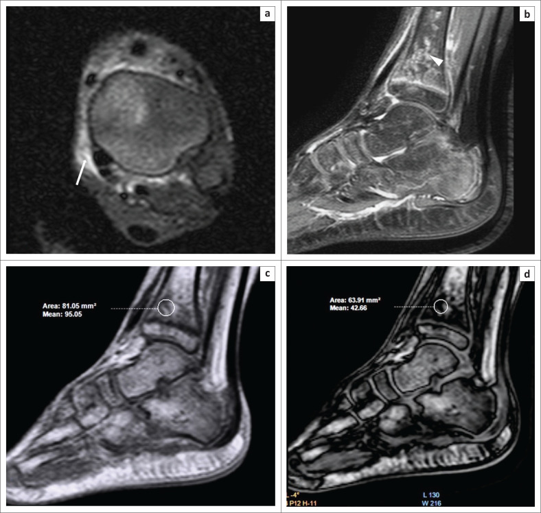
FIGURE 6.
Differentiating islands of red marrow from other sinister pathology. A 10-year-old boy presented with a 10-day history of pain and swelling over the left ankle. Magnetic resonance imaging showed soft tissue oedema (white arrow) over the medial aspect of the tibia on the axial Proton-Density-Weighted (PDW) spectral attenuated inversion recovery (SPAIR) image (a). Areas of hyperintensity were observed on PDW SPAIR (b) within the epiphysis and metaphysis of the distal tibia (white arrowhead). On comparing the in-phase (c) and opposed-phase (d) images, it was determined that the decrease in signal intensity on the opposed-phase images was 55% (>20%), which suggested the presence of red marrow and ruled out any sinister intraosseous pathological lesions such as osteomyelitis.

