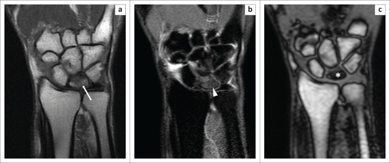
FIGURE 8.
Kienbock’s disease. A 32-year-old man underwent magnetic resonance imaging of the left wrist for the evaluation of chronic pain. The T1W image (a) revealed hypointensity (white arrow) within the lunate; the Proton-Density-Weighted spectral attenuated inversion recovery (b) image indicated hyperintensity (white arrowhead), which was suggestive of marrow oedema. The opposed-phase image (c) revealed a clear flattening of the lunate bone, with the sclerosed bone (white asterisk) demonstrating considerable hypointensity.

