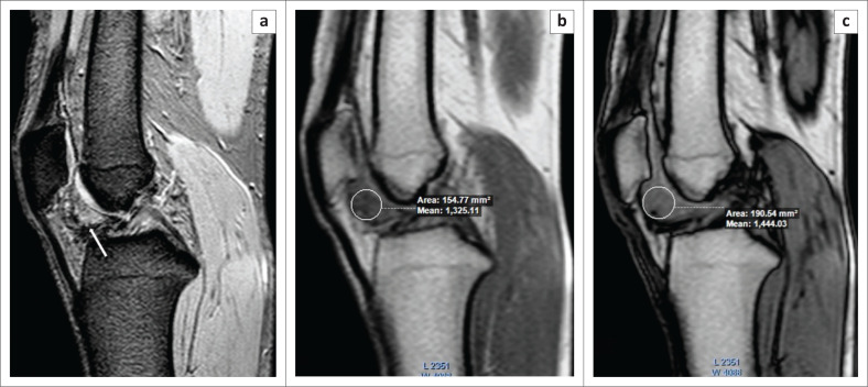
FIGURE 9.
Pigmented villonodular synovitis. A 19-year-old male presented with a history of right knee pain. The T2 GRE (Gradient Echo) image (a) showed hyperintense nodular soft tissue in Hoffa’s fat pad with a few areas of peripheral blooming on the GRE image (white arrow). At chemical shift imaging, the signal intensity of the nodular soft tissue was lower on the in-phase image (b) compared to the opposed-phase image (c), confirming the diagnosis of nodular pigmented villonodular synovitis.

