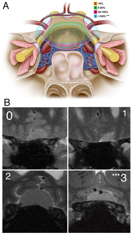
FIG. 1.
Optic nerve laterality score. A: The optic nerve laterality score was determined based on the maximal lateral extension of the tumor on the anterior skull base relative to the optic nerve on either side. We assigned a score of 0 if the maximal lateral extension on either side was medial to the optic nerve; 1 if lateral < 50%; 2 if lateral ≥ 50% but < 100%; and 3 if it was completely (≥ 100%) lateral to the optic nerve. B: The optic nerve laterality score was measured on coronal MRI. The coronal image demonstrates lateral extension above the optic nerve at the bony edge of the optic canal on the anterior skull base. ***Optic nerve laterality score of 3 was associated with a significantly increased risk of not achieving GTR. Panel A: Copyright Matthew Holt. Published with permission.

