Figure 2.

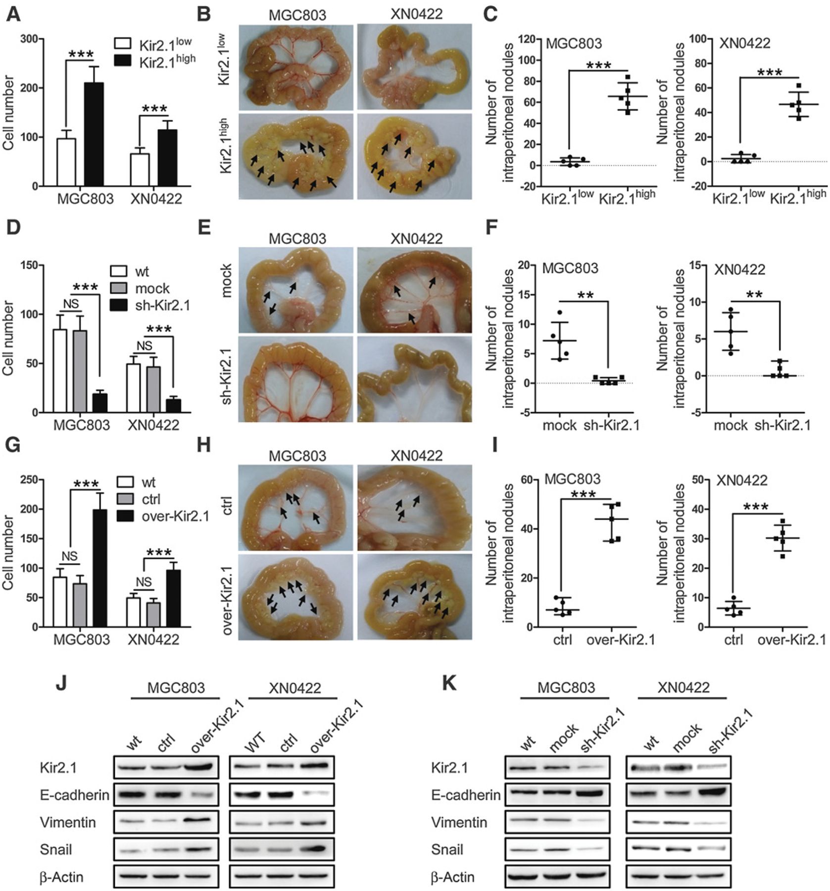
Kir2.1 promotes the invasion and metastasis of gastric cancer cells. A, Matrigel–Transwell invasion assay showing increased invasion capability of Kir2.1high gastric cancer cells compared with Kir2.1low gastric cancer cells. Data are shown as mean ± SD (n = 5; ***, P < 0.0001, Student t test). B, Representative images of intraperitoneal metastasis in NOD/SCID mice showing significantly higher number of metastatic foci formed by Kir2.1high gastric cancer cells than that formed by Kir2.1low gastric cancer cells. C, Quantification of metastatic tumors formed by Kir2.1high and Kir2.1low gastric cancer cells. Data are shown as mean ± SD (n = 5; ***, P < 0.0001, Student t test). D, Reduced invasion capability of sh-Kir2.1 gastric cancer cells compared with mock (scrambled shRNA) gastric cancer cells in vitro. Data are shown as mean ± SD (n = 5; NS, not significant; ***, P < 0.0001, ANOVA test). E and F, Reduced metastasis capability of sh-Kir2.1 gastric cancer cells in vivo. Data are shown as mean ± SD (n = 5; **, P < 0.001, Student t test). G, Increased invasion capability of over-Kir2.1 gastric cancer cells compared with control (empty vector) gastric cancer cells in vitro. Data are shown as mean ± SD (n = 5; NS, not significant; ***, P < 0.0001, ANOVA test). H and I, Increased metastasis capability of over-Kir2.1 gastric cancer cells compared with control (empty vector) gastric cancer cells in vivo. Data are shown as mean ± SD (n = 5; ***, P < 0.0001, Student t test). J, Western blotting showing decreased E-cadherin expression but enhanced vimemtin and Snail expression in over-Kir2.1 gastric cancer cells. K, Western blotting showing upregulated E-cadherin and downregulated vimemtin and Snail in sh-Kir2.1 gastric cancer cells.
