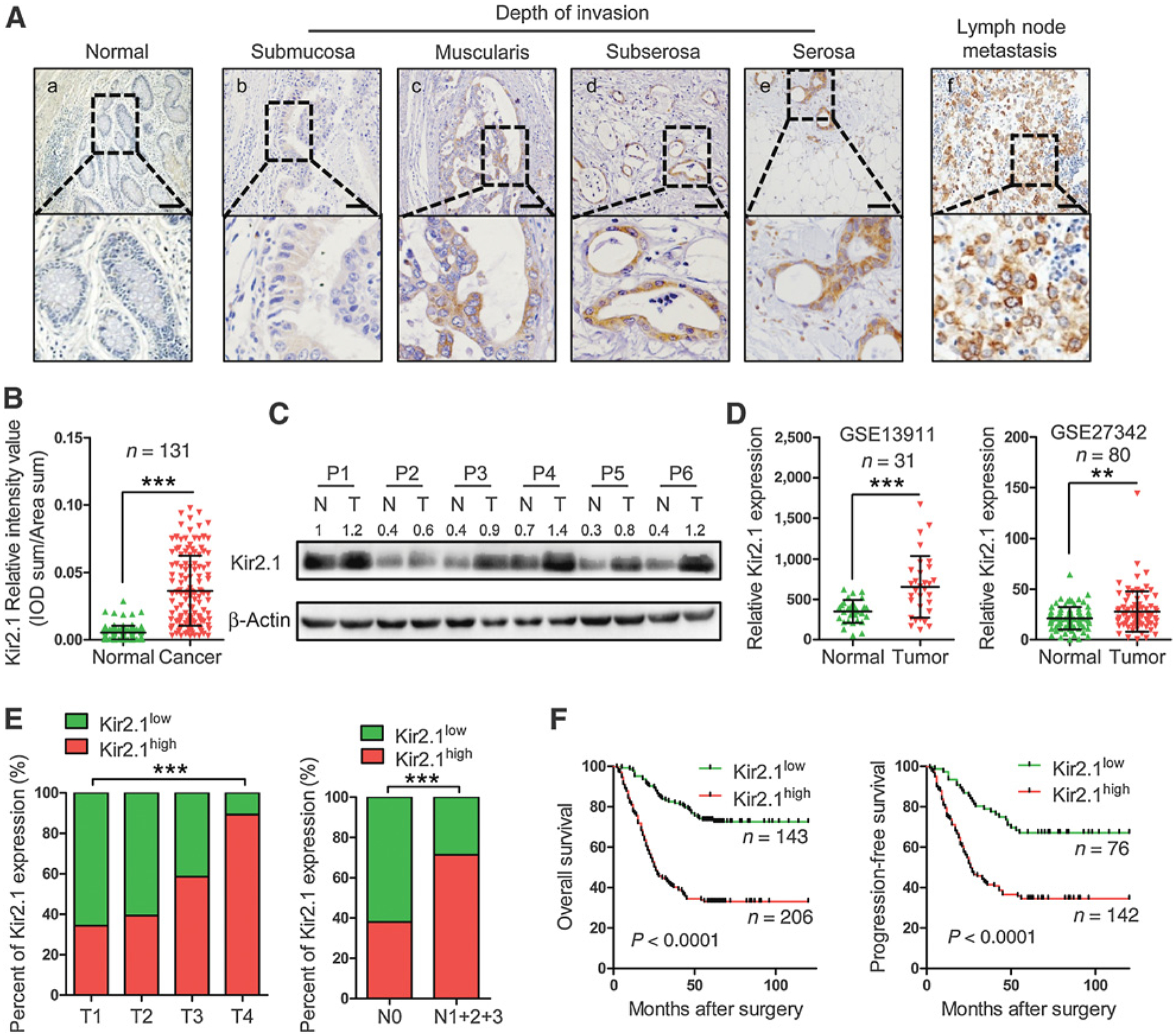
Figure 6.

Upregulation of Kir2.1 in human gastric cancer and the correlation with cancer invasion, lymph node metastasis, and outcome of patients with gastric cancer. A, Representative IHC images of Kir2.1 in gastric cancer specimens. Scale bar, 100 μm. a, Absence of Kir2.1 staining in normal gastric mucosa. b–e, Increased Kir2.1 staining intensity with invasion depth. f, High Kir2.1 level in both primary tumor and corresponding metastatic lymph node. B, Higher IHC scores of Kir2.1 in 131 gastric cancer tissues compared with paired adjacent normal tissues. Data are shown as mean ± SD (n = 131; ***, P < 0.0001, paired t test). C, Kir2.1 protein in 6 paired surgically removed gastric tumor tissues (T) and the adjacent normal tissues (N) showing highly expressed Kir2.1 in cancerous tissues. D, Higher mRNA level of Kir2.1 in cancerous tissues than in adjacent normal tissues from GEO GES13911 and GES27342. Data are shown as mean ± SD (n = 31, GSE13911; n = 80, GSE27342; ***, P < 0.0001; **, P < 0.001, paired t test). E, The relationship between Kir2.1 expression and tumor invasion depth and lymph node metastasis of gastric cancer. n = 349; ***, P < 0.0001, χ2 test. F, Kaplan–Meier curves showing the correlation between the levels of Kir2.1 and the overall survival and progression-free survival of patients with gastric cancer [n = 349 (OS), n = 218 (PFS), log-rank test].
