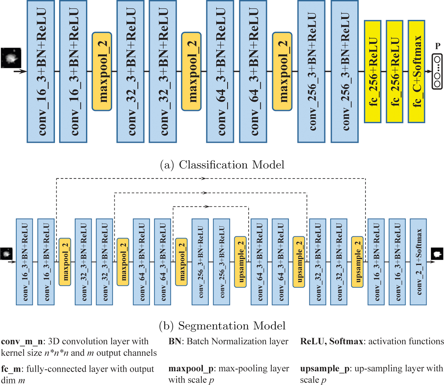
Figure 2:

The classification model (a) is 3D CNN containing 8 convolution blocks, 3 max-pooling layers and 3 fully-connected blocks. The segmentation model (b) is a 3D U-Net with 15 convolution blocks, 3 max-pooling layers and 3 up-sampling layers. Dilated-convolution is exploited in the segmentation model to increase the receptive field.
