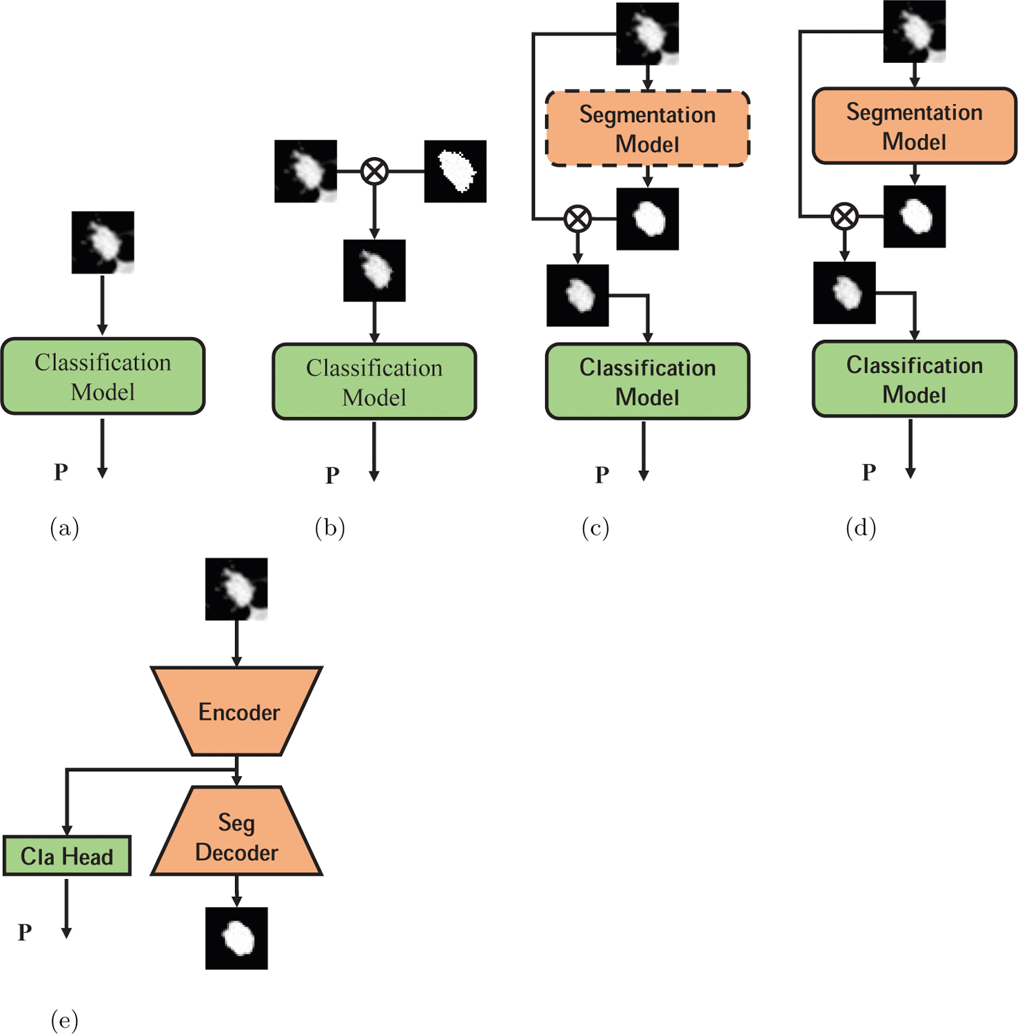
Figure 3:

Different deep learning models used in this paper. (a) Train classification model with raw CT data. (b) Train classification model with data masked by ground-truth segmentation. (c) Train segmentation model first, then train classification model with data masked by automatically generated segmentation map, dash rectangle means the model parameters are fixed. (d) Jointly train classification model and segmentation model. (e) Multi-task learning model with segmentation and classification.
