Figure 1.
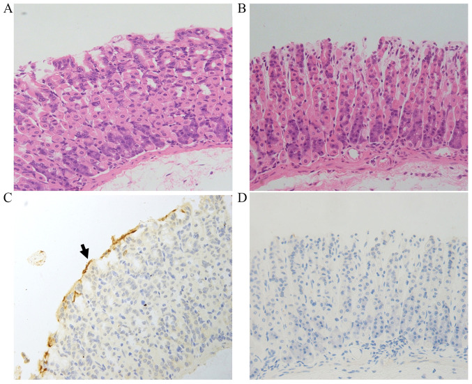
HP bacterial colonization in gastric tissue. Hematoxylin and eosin staining of gastric tissue sections from the (A) HP+ and (B) HP- groups. Immunohistochemical staining of HP in gastric tissue sections from the (C) HP+ and (D) HP- groups. The arrow indicates the HP bacterial colony. Magnification, x400. HP, Helicobacter pylori.

