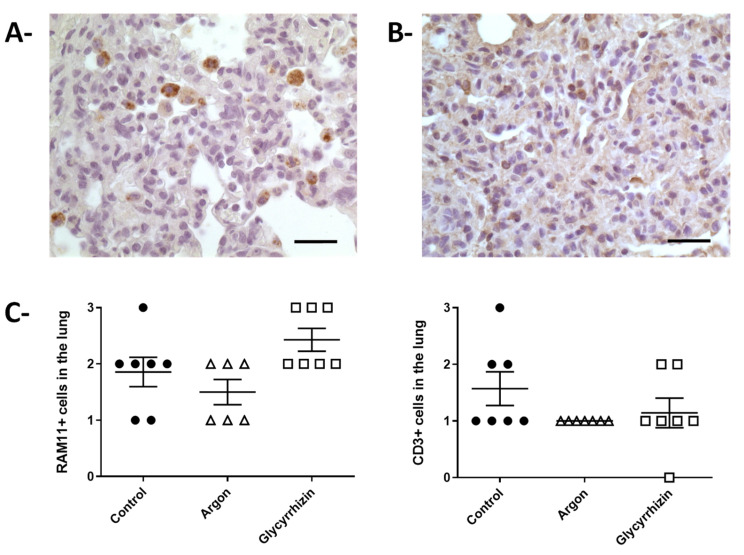
Figure 6.
Immunochemistry in lung parenchyma. (A)—Morphological appearance of the immunohistochemical staining for macrophage identification using the RAM11 polyclonal antibody (bar = 25 µm). (B)—Morphological appearance of the immunohistochemical staining for T cells identification using a CD3 antibody (bar = 25 µm). (C)—Semiquantification of RAM11 and CD3 positive cells in the pulmonary samples from the different groups.

