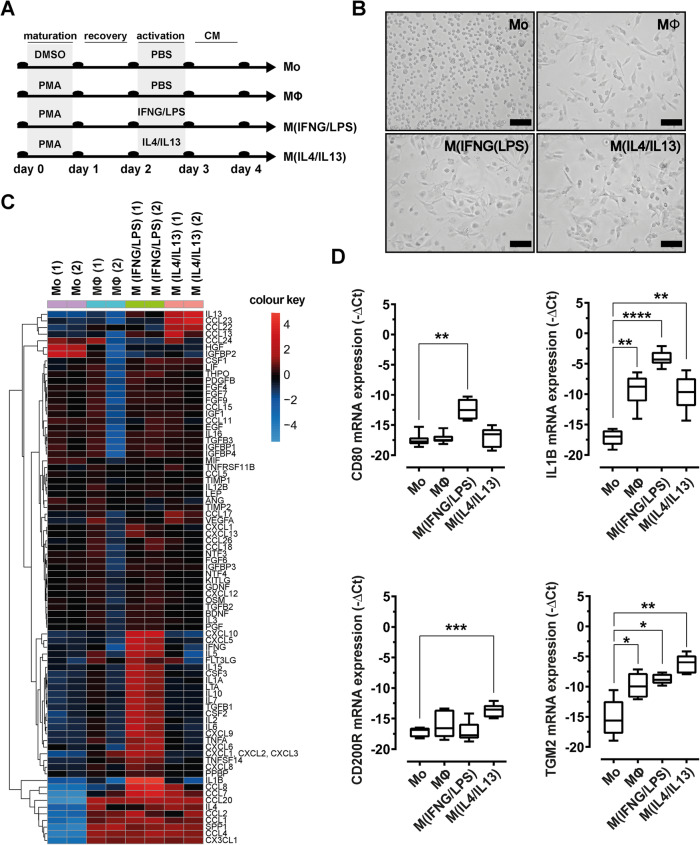
Fig. 2.
Characterization of the macrophage activation model. a THP1 cell treatment protocol for in vitro polarization of macrophages. b Morphology (phase contrast, scale bar 100μm) of THP1-derived macrophages on day of CM harvest. c Heatmap of log2 cytokine protein levels in differentially activated macrophages. The dendrogram represents the result from a complete linkage hierarchical clustering of mean-centered expression levels based on Euclidian similarity. d mRNA-expression levels of macrophage-selective markers (M(IFNG/LPS): CD80, IL1B, and M(IL4/IL13): CD200R, TGM2). Data were normalized to the 18S rRNA reference gene and shown as mean –ΔCt. Asterisks indicate p values < 0.05 (*), p<0.01 (**), and p<0.001 (***). Statistical significance (n=5) was determined by using one-way ANOVA and Dunnett’s test for multiple comparison

