Fig. 1.
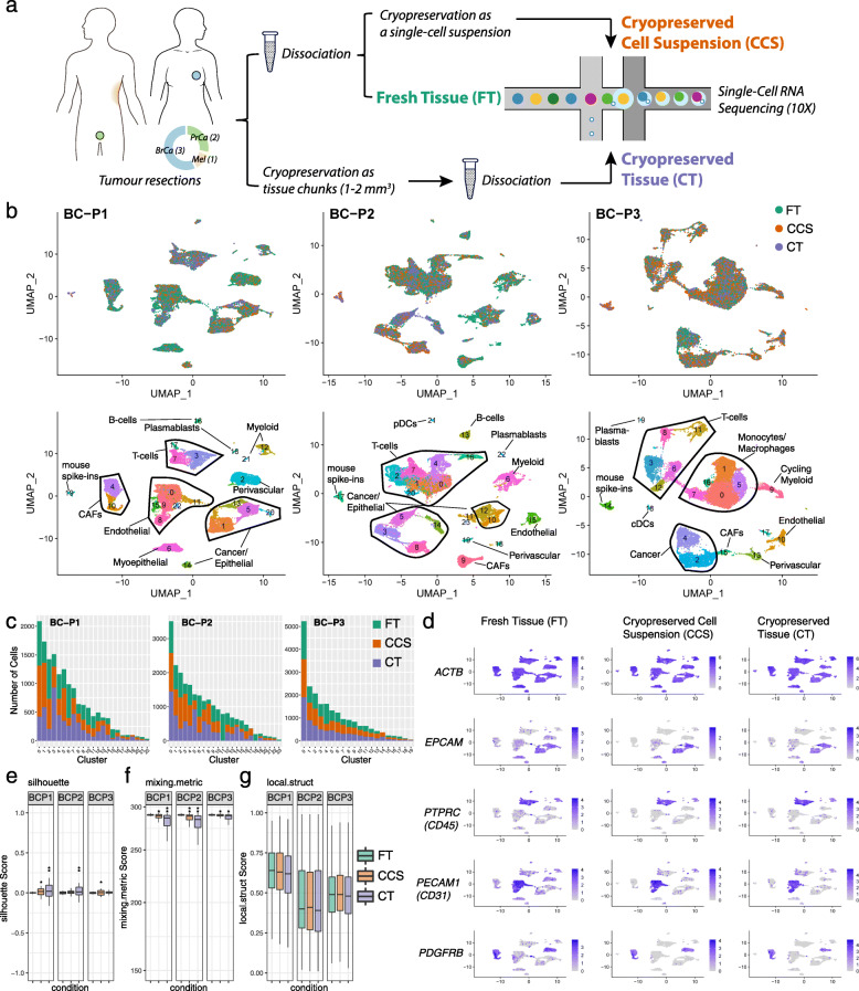
Cryopreservation allows for robust cell-type detection in clinical breast cancer samples. a Experimental workflow. b UMAP visualisation of 23,803, 29,828 and 24,250 cells sequenced across dissociated fresh tissue (FT; green), dissociated cryopreserved cell suspensions (CCS; orange) and solid cryopreserved tissue (CT; purple) replicates from three primary breast cancer cases (BC-P1, BC-P2 and BC-P3). UMAPs are coloured by cryopreserved replicate (top) and by cluster ID (bottom) with cell types annotations overlayed. Matched replicates were integrated using the Seurat v3 method. c Number of cells detected per cluster. Cells were downsampled to the lowest replicate size. d FeaturePlot visualisations of gene expression from BC-P1 fresh and cryopreserved replicates, showing the conservation of the housekeeping gene ACTB and heterogeneous cancer/epithelial (EPCAM), immune (PTPRC/CD45), endothelial (PECAM1/CD31) and fibroblast/perivascular (PDGFRB) clusters. e, g Distribution of silhouette scores (range −1 to + 1) (e), mixing metric (f) and local structure metrics (g) of clustering following cryopreservation. Samples were downsampled by replicate and cluster sizes and compared to the respective FT samples. Cell comparisons were performed across downsampled FT-1 vs FT-2 cells (positive control), FT vs CCS cells and FT vs CT cells. Stars represent standard deviations: e silhouette scores s.d. 0.02–0.05* and s.d. > 0.05**; f mixing metrics s.d. 2–10* and s.d. > 10**; g local structure metrics s.d. > 0.05*

