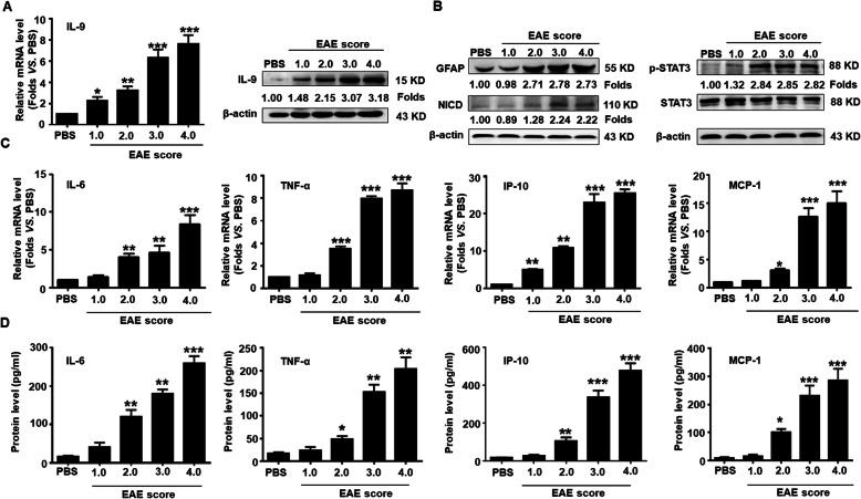
Fig. 1.
Upregulation of IL-9 and inflammatory cytokines as well as the activation of Notch1 signaling during EAE process. a The level of IL-9 in spinal cords of EAE mice with different clinical scores was measured by real-time PCR and Western blot assay, respectively. b The expressions of GFAP, NICD and p-STAT3 in spinal cords along with EAE process were detected by Western blot assay. c The mRNA levels of IL-6, TNF-α, IP-10, and MCP-1 in spinal cords were determined using real-time PCR. d The secretion levels of IL-6, TNF-α, IP-10 and MCP-1 in the sera were detected by Cytometric Bead Array (CBA) during the EAE process. *p < 0.05, **p < 0.01 and ***p < 0.001 versus PBS group (n = 6/group, one-way ANOVA). These results were repeated four times. Data are represented as the mean ± SEM

