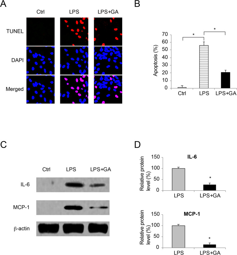
Fig. 2.
The effect of GA on LPS-induced WI-38 apoptosis and inflammation.aWI-38 cells were cultured in vitro (Ctrl), or treated with 10mg/ml LPS for 24h (LPS), or pre-incubated with 50 nM GA for 24h prior to LPS treatment (LPS + GA). A TUNEL assay was applied to identify apoptotic cells with TUNEL (Red) immunoreaction. During the meantime, DAPI (Blue) immunoreaction was applied to identify WI-38 cell nuclei. bFor images acquired in TUNEL assay, the percentages of apoptotic WI-38 cells were compared among Ctrl, LPS and LPS + GA conditions (* P < 0.05). cFor WI-38 cells under Ctrl, LPS and LPS + GA conditions, western blot analysis were conducted to compare IL-6 and MCP-1 protein expressions. dFor western blot data in (c), relative band intensities of IL-6 and MCP-1 were compared between LPS and LPS + GA conditions (* P < 0.05)

