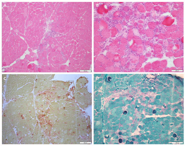
Figure 1.

(A) H&E which shows perimysial perivascular mononuclear inflammatory cell infiltrates at 10× magnification. (B) H&E which shows necrotic and basophilic fibres at 40× magnification. (C) Gomori trichrome stain which shows necrotic and basophilic fibres at 10× magnification. (D) Acid phosphatase stain which shows the necrotic fibres staining red at 4× magnification; the necrotic fibres have faint pink cytoplasm. Images are courtesy of the Neuromuscular Laboratory at the University of Rochester
