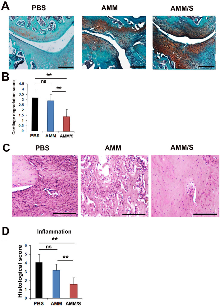
Fig. 5. Histological staining of joints in CIA mice after injection of cells.
(A) Proteoglycan expression was identified using Safranin O staining of the joints of CIA mice after injection of MSCs. Scale bars = 200 μm. (B) Quantification of cartilage degradation scores. Loss of proteoglycans was identified after staining for proteoglycans. n = 5 each; **P < 0.01. ns, not significant. (C) Representative images of H&E-stained sections of joint tissues. Scale bars = 200 μm. (D) Quantification of inflammatory response histological scores. Mononuclear cell infiltration and inflammatory pathological scores were measured after cell transplantation. AMM/S-injected Treg and Th17 cells exhibited low mononuclear cell infiltration and normal cartilage surface morphology. n = 5 each; **P < 0.01.

