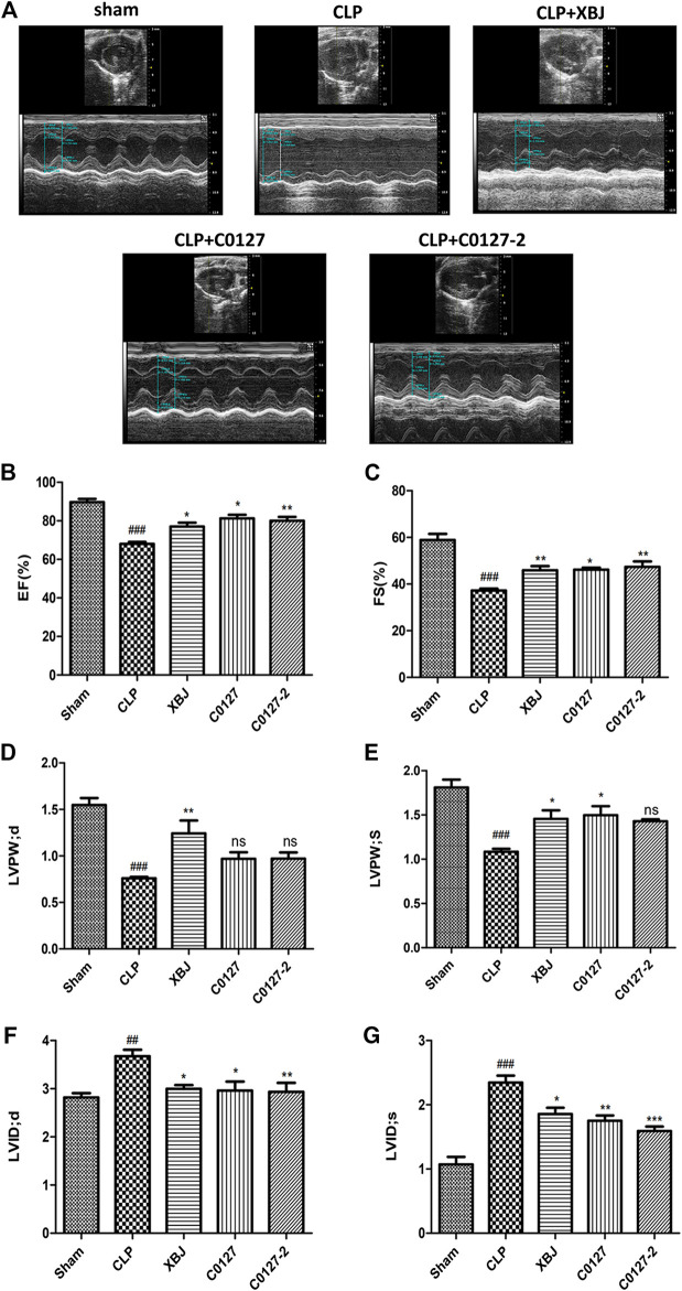
FIGURE 2.
XBJ and key compounds in XBJ protected cardiac function of septic mice. (A) Representative images of left ventricular echocardiography. Cardiac performance was determined by echocardiography in different groups as indicated. (B) Left ventricularejection fraction (LVEF) %, (C) Left ventricular fractional shortening (LVFS) % were measured in M-mode, (D) LV posterior wall diastole (LVPWd), (E) LV posterior wall systole (LVPWs), (F) Left ventricular internal dimensions at diastole (LVIDd), (G) Left ventricular internal diameter systole (LVIDs), Results were presented as mean ± SEM (n = 7–10/group). # p < 0.05, ## p < 0.01, ### p < 0.001 vs. Sham group, *p < 0.05, **p < 0.01 vs. CLP group.

