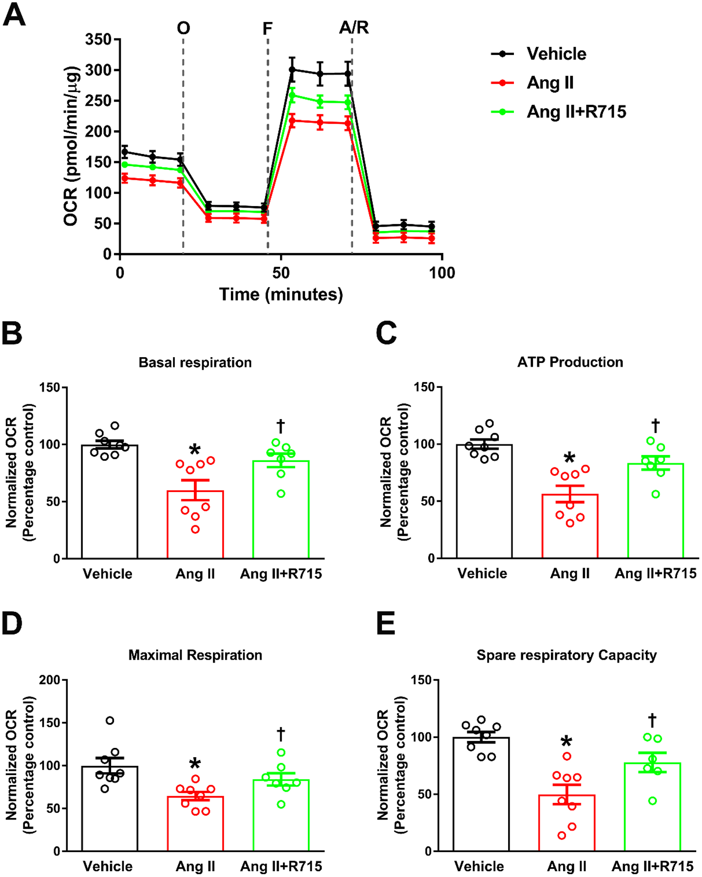
Fig. 5. B1R antagonist treatment prevented angiotensin II-induced decrease in mitochondrial respiration in primary hypothalamic neurons.

Primary hypothalamic mouse neurons were cultured on Seahorse XF-24 plates and treated with vehicle or Ang II with or without R715 for 24 hours. Seahorse mito stress assay was performed to measure oxygen consumption rate (OCR). (A). Bioenergetic profile following sequential injection of oligomycin (O), FCCP (F) and antimycin A/rotenone (A/R) indicating the key parameters of mitochondrial respiration in neurons. OCR measurements showed that Ang II stimulation resulted in significant decrease in basal respiration (B), ATP production (C), maximal respiration (D), and spare respiratory capacity (E) indicating mitochondrial dysfunction, and treatment with R715 prevented this Ang II-induced mitochondrial dysfunction. (n=8 independent culture wells/group). Statistical significance: One-way ANOVA followed by Tukey’s multiple comparisons test. *p<0.05 compared to vehicle, †p<0.05 compared to Ang II.
