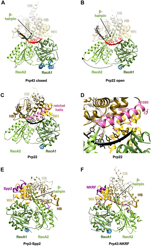
Figure 2.
Structural Basis for the DEAH-Box Mechanism of Action and Regulation by G-Patch Domains.
The domains are colored as in Figure 1A. (A) The closed form of Chaetomium thermophilum DEAH-box helicase Prp43 bound to ADP and RNA (PDB ID 5LTA). (B) The open form of C. thermophilum DEAH-box helicase Prp22 bound to RNA (PDB ID 6I3P). Both proteins are shown in the same orientation after aligning their RecA1 domains. The RNA stacked in the binding tunnel is highlighted in red. (C) Location of the ratchet helix in C. thermophilum Prp22 (PDB ID 6I3P). The ratchet helix is highlighted in pink. (D) A zoomed in view of the ratchet helix and its surroundings. Residues on the ratchet helix that interact, or potentially interact, with a longer RNA are shown as sticks. (E) The structure of C. thermophilum Prp2 in complex with Spp2 (purple) (PDB ID 6RM9). (F) The structure of Homo sapiens Prp43 with NKRF (purple) bound (PDB ID 6SH7). Abbreviations: HB, helix bundle; OB, oligosaccharide-binding fold; WH, winged helix.

