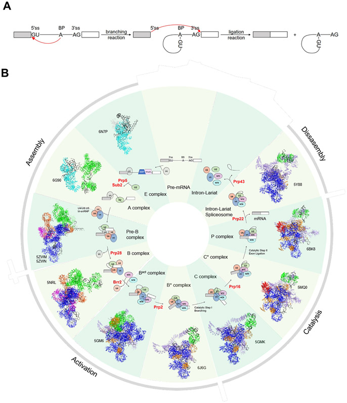
Pre-mRNA Splicing. (A) Schematic representation of the two transesterification reactions in pre-mRNA splicing. Boxes and solid lines represent the exons and introns, respectively. The red arrows show the nucleophilic attacks at the phosphodiester bond at the 5′ and 3′ ss during splicing. (B) A schematic representation of the splicing cycle in yeast is shown in the inside ring. Only the snRNPs (ovals) but not non-snRNP proteins are shown for simplicity. The spliceosomal helicases are indicated in red. The outside ring shows the cryo-EM structure of each corresponding spliceosomal complex and its PDB ID.

An official website of the United States government
Here's how you know
Official websites use .gov
A
.gov website belongs to an official
government organization in the United States.
Secure .gov websites use HTTPS
A lock (
) or https:// means you've safely
connected to the .gov website. Share sensitive
information only on official, secure websites.
