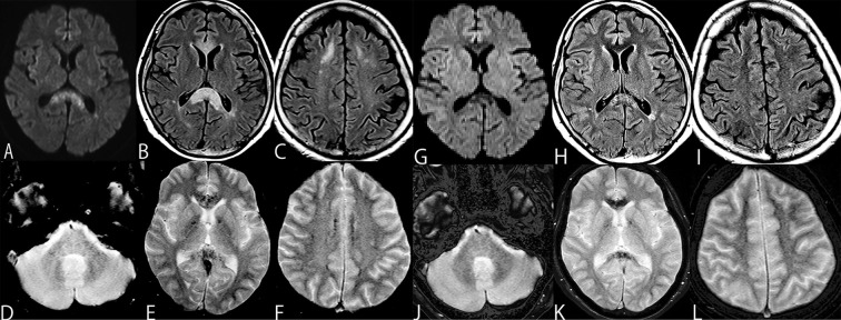
Figure 1.
MRI findings on the 12th day, DWI and FLAIR showed widespread areas of high intensity in the splenium of the corpus callosum (A, B) and white matter in the centrum semiovale (C). T2* revealed extensive microbleeds that were symmetrically located in the middle cerebral peduncle (D) and anterior to the splenium of the corpus callosum and globus pallidus (E), and the white matter in the centrum semiovale (F). At 41 days after the onset of symptoms, DWI and FLAIR showed improvement in the high intensity lesions in the corpus callosum and the splenium lesion (G, H), as well as the disappearance of the white matter lesions (I). The widespread and marked appearance of microbleeds was decreased; however, some microbleeds remained (J-L).

