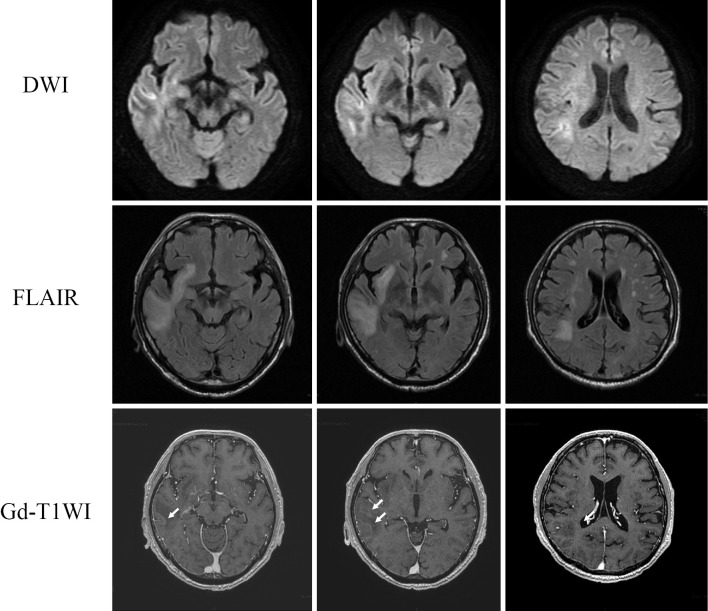
Figure 1.
MRI findings on day 3 of admission during steroid pulse therapy. A high signal in the cortex and white matter of the right temporal lobe on DWI, FLAIR images with gadolinium enhancement at the margins of the lesion in the right temporal lobe (arrows). DWI: diffusion-weighted imaging, FLAIR: fluid-attenuated inversion-recovery, T1WI: T1-weighted imaging, MRI: magnetic resonance imaging, Gd: gadolinium

