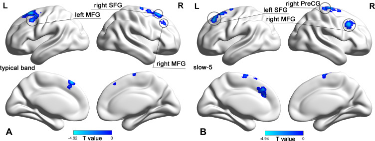
Figure 2.
dfALFF map of non-NPSLE patients versus healthy controls in different frequency bands T-statistic dfALFF maps of non-NPSLE patients versus healthy controls (HCs) (voxel p<0.005, cluster p<0.05, GRF corrected). (A) dfALFF map of non-NPSLE patients versus healthy controls in typical band; (B) dfALFF map of non-NPSLE patients versus healthy controls in slow-5. Hot colors indicate increased dfALFF in patients compared with HCs; cold colors indicate decreased dfALFF in patients compared with HCs.
Abbreviations: R, right hemisphere; L, left hemisphere; MFG, middle frontal gyrus; SFG, superior frontal gyrus; PreCG, precentral gyrus.

