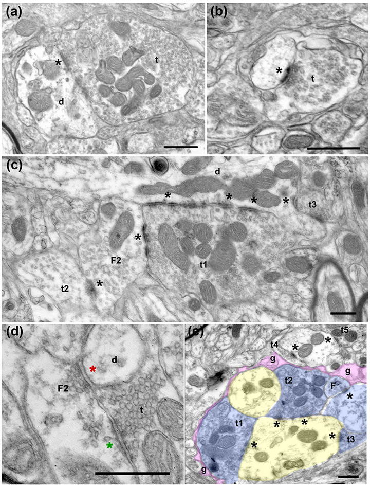
Figure 3.
Electron micrographs of terminals in the tree shrew VPMP. (a) A large terminal (t) filled with round vesicles and many mitochondria makes a synapse onto a large dendrite (d). The asterisk (*) marks the synapse at the postsynaptic side, here and in all subsequent panels. (b) A small terminal (t) with sparse vesicles synapses onto a dendrite. (c) A large terminal (t1) filled with round vesicles and many mitochondria makes a synapse onto a dendrite (d) and a vesicle filled profile (F2; classified as such because it contains pleomorphic-vesicles and it is postsynaptic to another terminal). Note that both synapses display multiple active zones (*). Another terminal (t2) with sparse pleomorphic vesicles (presumed inhibitory) synapses onto the same F2 profile, representing an inhibitory-to- inhibitory interaction. A third terminal (t3) that is notably small compared to t1, synapses onto the large dendrite. (d) A terminal (t) with round vesicles synapses onto a presynaptic dendrite (F2). The green asterisk marks the putative excitatory synapse. The presynaptic dendrite (F2), in turn, forms a symmetrical, presumed inhibitory synapse (red asterisk) onto another dendrite (d). (e) In a glomerular structure, three terminals (t1, t2, t3) that are pseudo-colored in blue form synapses onto a large dendrite (yellow). The synaptic arrangement is encased in an astrocytic sheath (g, pink). The synapses are indicated by asterisks at the postsynaptic side. Scale bars= 500 nm.

