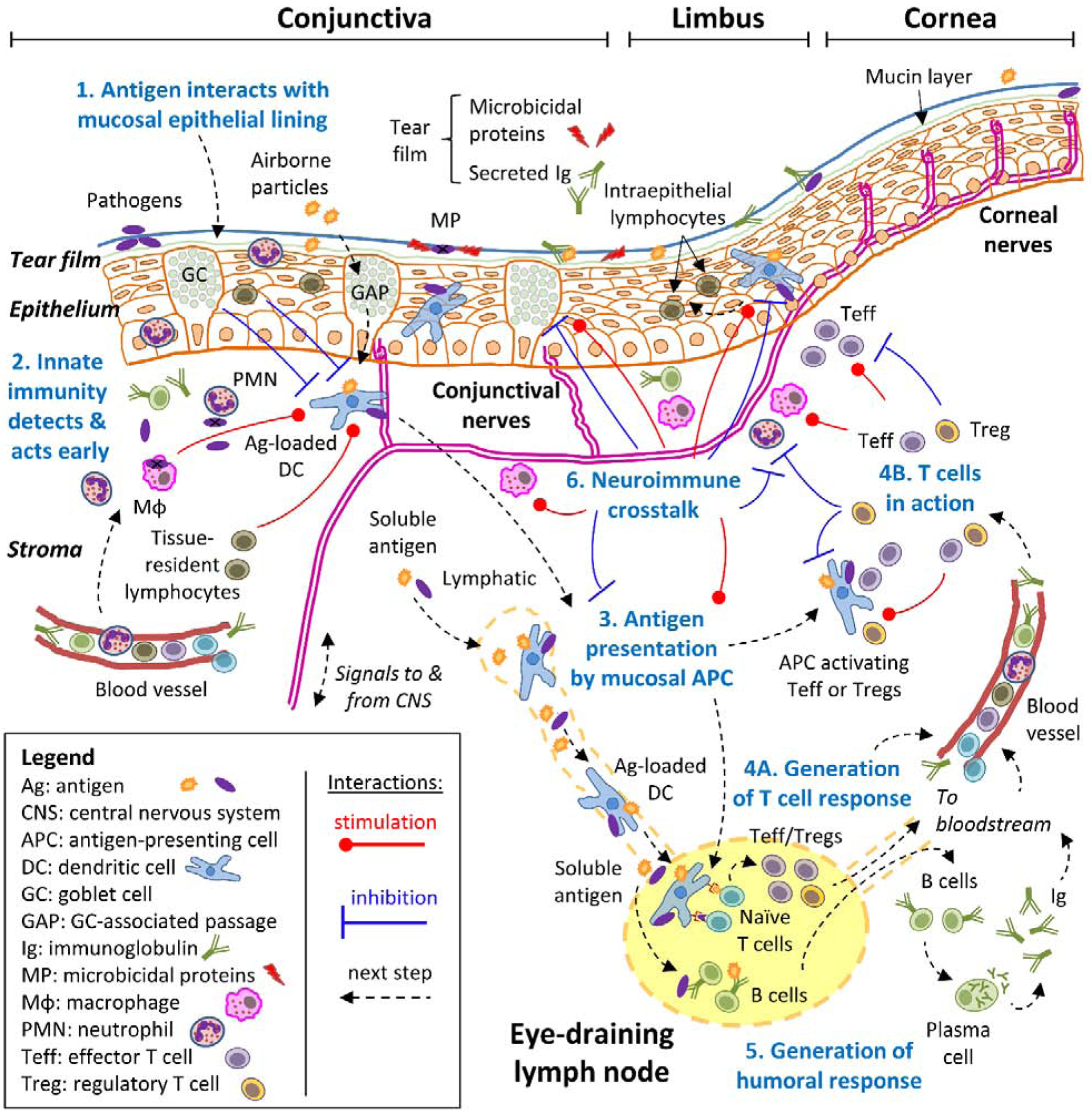
Figure 1: Schematic mucosal immune response of the ocular surface.

A simplified approach to the mucosal immune response to diverse antigens, consisting of 6 consecutive steps (numbered in blue), is exemplified and adopted throughout this review. For clarity, all steps revolve around the concept of an antigen (the molecule or molecular structure against which the adaptive immune system reacts), which can derive from pathogens, foreign substances, or even self-tissues. Step 1: the antigen interacts with the mucosal epithelial lining, which acts as a barrier aided by blinking action and the tears. Step 2: antigens may breach through the epithelial barrier, setting off the innate immune system’s detection systems because receptors on diverse cell types recognize molecular patterns that are associated with potentially dangerous entities. This triggers a swift defense that results in early containment and that influences the next step at the same time. Step 3: specialized antigen-presenting cells (APC) that reside in the ocular surface capture antigens and migrate to the eye-draining lymph nodes, where they process them and present antigen-derived peptides on their surface. Step 4: naïve T cells that continuously recirculate through lymph nodes recognize specific peptides on APC, become activated, and then expand and differentiate to either effector (Teff) or regulatory (Treg) T cells depending on additional signals that they pick up during antigen presentation. These antigen-experienced T cells leave the lymph node and through the bloodstream, reach the ocular surface where they can exert their many functions in the immune response. Step 5: concomitantly with step 3 and 4, soluble antigens reach the lymph node from the ocular surface, where B cells might interact with them, become activated, and then expand and differentiate into plasma cells that migrate to other tissues through the bloodstream, mainly the bone marrow. Once at their final destination, plasma cells secrete large amounts of immunoglobulin (Ig) that reach the ocular surface and the lacrimal glands through the bloodstream. Step 6: the nervous system can also sense signals associated with some antigens and interacts with every cell type of the immune response in a bidirectional process (neuroimmune regulation). Although most of the immune response takes place at the ocular surface, it should be noted that critical steps 3, 4, and 5 begin in the draining lymph node. Also, the immune system is continuously interacting with various antigens, and thus, these processes take place simultaneously.
