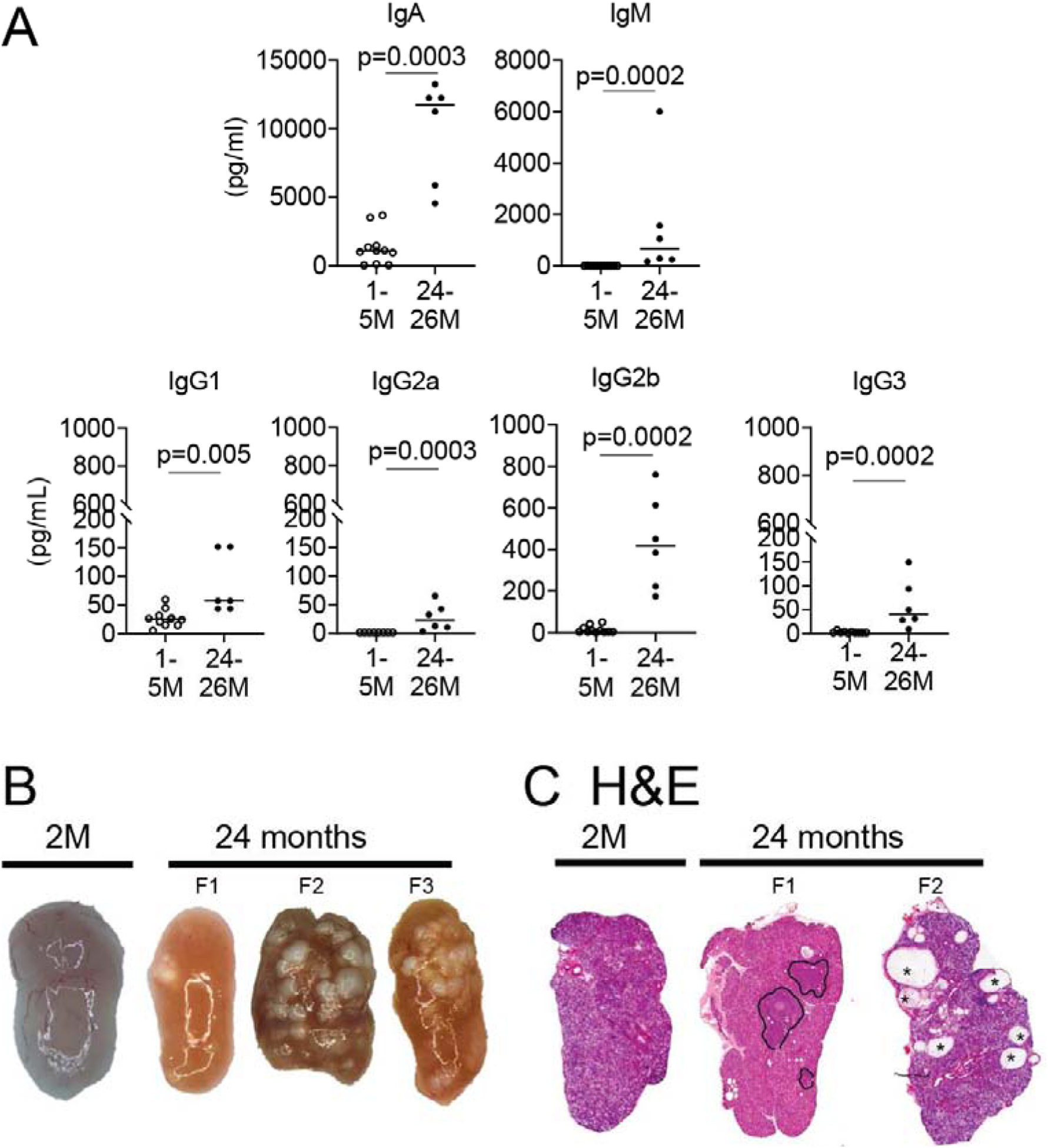
Figure. 2. Tear film and lacrimal gland changes during aging.

A. Tear washings from young and aged mice of both sexes were collected, and immunoglobulins were measured using Luminex assay. Each dot corresponds to tear washings from 10 animals (20 eyes). B. Representative macro images of young and aged female lacrimal glands. Note the yellowish-tan appearance of aged lacrimal gland and presence of cysts (present in 20–30% of aged lacrimal glands, F2 and F3). C. Representative scans of lacrimal gland sections stained with H&E from young and aged B6 female mice. Areas of lymphocytic infiltration are demarcated aged glands (F1), and areas of dilated ducts/cysts are easily identified (F2, asterisks). 2M = 2 months; F = female.
