
Figure 1.
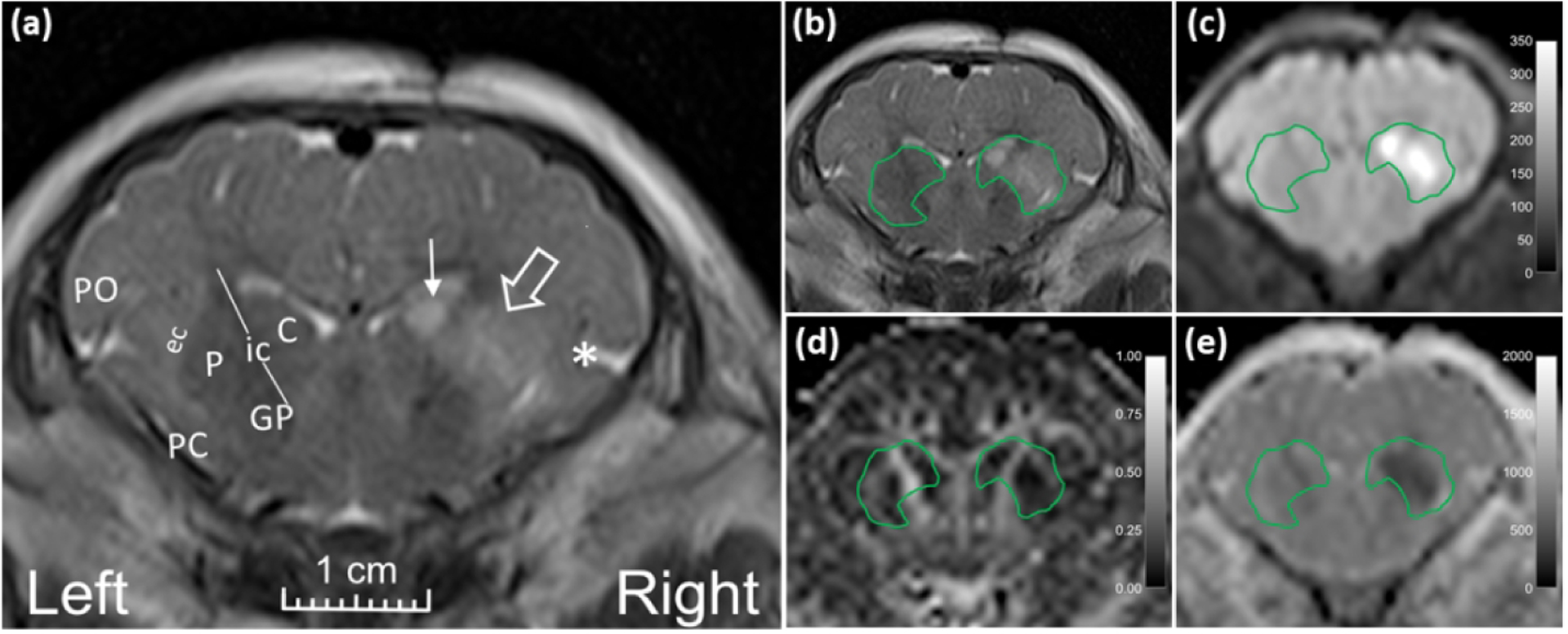
Representative MR images from a neonatal piglet that received stereotaxic injections of quinolinic acid (QA; 960 nmol, right) and contralateral phosphate-buffered saline (PBS, control, left) with 24-h survival. (a) High signal in the putamen (open arrow), caudate (line arrow), and deep insular cortex (asterisk) on the QA-injected side is visible on the T2-weighted coronal image. Anatomic landmarks are shown in the PBS-injected side: P, putamen; C, caudate; ic, internal capsule; GP, globus pallidus; ec, external capsule; PC, piriform cortex; and PO, parietal operculum. Manually defined regions of interest (ROI) encompassing the putamen, internal capsule, and caudate in (b) T2-weighted and (c) diffusion-weighted images show increased signal from QA injury. (d) Fractional anisotropy and (e) mean diffusivity were measured in the ROIs containing putamen, internal capsule, and caudate.