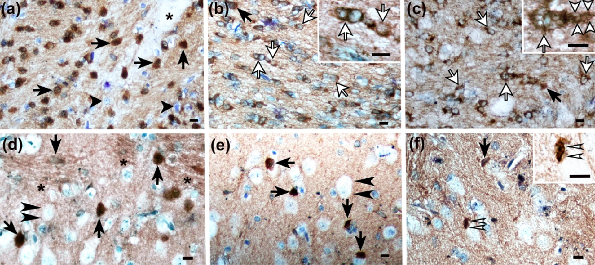
Figure 10.

Immunophenotyping and classification of normal and injured mature oligodendrocytes in piglet striatum with bridging integrator-1 (BIN1). (a) Internal capsule of a sham piglet showed normal oligodendrocytes with BIN1 immunoreactivity in the nucleus and cytoplasm (arrows). Normal BIN1-negative cells are identified by arrowheads. A neuron is marked by an asterisk. (b) The internal capsule of a piglet with hypoxia-ischemia had degenerating BIN1+ oligodendrocytes that exhibited vacuoles in the cell soma and nuclear exclusion of BIN1 (white arrows). The inset shows vacuoles and loss of BIN1 immunoreactivity. The black arrow indicates a normal BIN1+ oligodendrocyte that expresses BIN1 within the normal-appearing nucleus and cytoplasm. (c) In pigs that were injected with 960 nmol quinolinic acid (QA), multiple degenerating BIN1+ oligodendrocytes were identified as having vacuoles in the nucleus and cytoplasm alongside nuclear extrusion of BIN1 (white arrows). The arrowheads in the inset show vacuoles in the BIN1+ oligodendrocyte’s processes. The black arrow identifies a normal BIN1+ oligodendrocyte. (d) Putamen in a sham piglet had normal BIN1+ oligodendrocytes (arrows). Neurons were not immunoreactive for BIN1 (double arrowheads). Each microscope field was randomly placed in putamen and included predominantly gray matter parenchyma plus some white matter bundles (asterisks). BIN1+ oligodendrocytes were counted in gray and white matter parenchyma within the putamen microscope field. (e) A sham piglet’s caudate had normal BIN1+ oligodendrocytes (arrows) and BIN1-negative neurons (double arrowheads). (f) An apoptotic BIN1+ cell (double white arrows) and normal BIN1+ oligodendrocyte (arrow) in caudate of a piglet injected with 960 nmol QA. The apoptotic, karyorrhectic nuclear fragmentation is visible in the inset. Main panels were photographed at 400x. Insets B, C, and F were photographed at 1000x with oil immersion. All scale bars are 10 μm.
